baseline vaginal ultrasound
Call 514 843-1650 on day 1 of your cycle to request an appointment. The baseline ultrasound should be done on day two or day three of your period.
![]()
A Transvaginal Ultrasound Image For One Pregnant Patient After Rat Download Scientific Diagram
These follicles could vary in size from 2-10 mm.

. First a clear gel is applied to the area of interest which improves the image quality of the study. During the exam you lie on a table or sit in a chair in one of our dedicated ultrasound rooms. Before initiating tamoxifen baseline vaginal ultrasound sonohysterography or office hysteroscopy to exclude the presence of endometrial polyps appears appropriate.
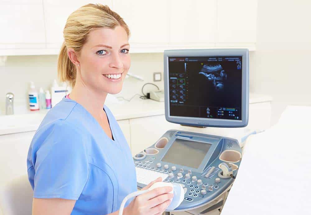
Pelvic organ ultrasound is used to monitor pregnancy find cysts on your ovaries examine the lining of your uterus look for causes of infertility and find cancers or benign tumors in the pelvic region. A transvaginal ultrasound can serve many purposes so the goal of yours depends on the type of ultrasound our Las Vegas fertility doctors prescribe. Iui infertility vaginalultrasoundWatch as I discuss what the baseline ultrasound process was like what the doctor is looking for and what to expect next.
Ultrasound examinations are performed by one of our highly trained staff of sonologists or board-certified radiologists at NYU Langone. A transvaginal ultrasound gives your fertility care team a look at your reproductive organs including your uterus and ovaries. The Basal Antral Follicle Count along with the womans age and Cycle Day 3 hormone levels are used as indicators for estimating ovarian reserve and the womans chances for pregnancy with.
The purpose is to check that there are no unusual cysts on the ovaries before starting the fertility drugs. The baseline vaginal ultrasound is performed at the. Obstetrics and Gynecology Ultrasound 6th floor of Block C Room C 064351.
At the same visit we will perform a baseline transvaginal ultrasound to measure the ovaries uterus and ovarian activity through an antral follicle count AFC. Antral follicle count is rightly done on day 3 of the cycle by a Trans vaginal ultrasound. Hysterosonography is performed very much like a gynecologic exam.
Look for cancer in your ovaries uterus or bladder. Depending on the view needed the ultrasound sensor is placed either on your abdomen pelvic ultrasound or in your vagina transvaginal. The ultrasound will take about 45 minutes to complete.
Some doctors use ultrasound during embryo transfer. Find an intrauterine device IUD. The baseline ultrasound for a frozen embryo transfer cycle.
Further the number of small antral follicles in both the ovaries is measured. A baseline scan is also done at the beginning of a frozen embryo transfer FET cycle. A requisition is needed if you are doing this ultrasound.
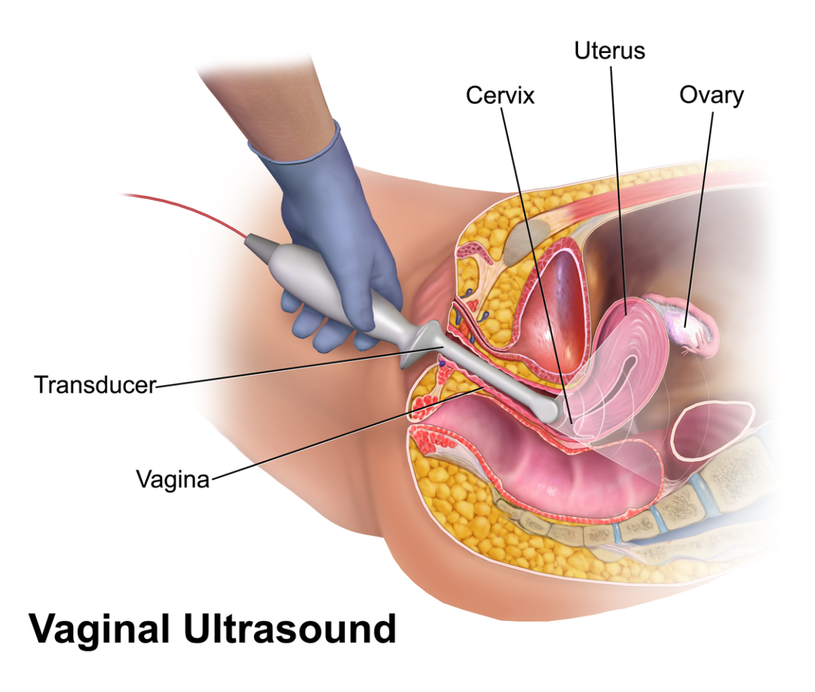
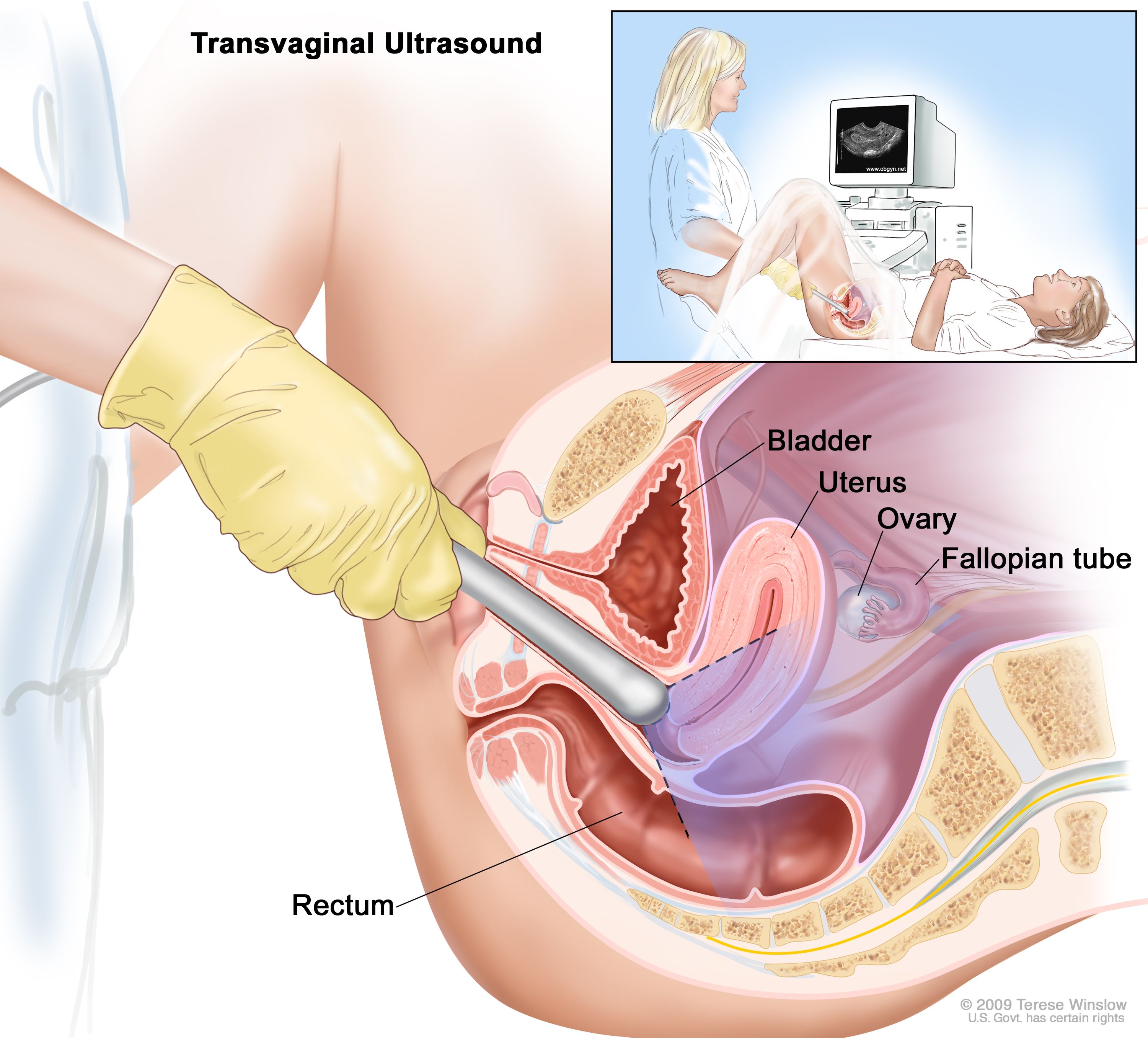
Ultrasound is also used during IVF for egg retrieval to guide the needle through the vaginal wall to the ovaries. The ultrasound is trans-vaginal a wand-like transducer is inserted vaginally to look at your ovaries uterus and endometrial lining. A probe is gently applied against the skin.
Day 3 testing consists of bloodwork and an ultrasound that are completed on the third day of a womans menstrual cycle. Initially the ovarian volume of both the ovaries is calculated. These tests ensure that you dont have any cysts in your ovaries the lining of your uterus is thin and to confirm that.
Find problems with the structure of your uterus or ovaries. Hysterosonography also called sonohysterography uses sound waves to produce pictures of the inside of a womans uterus and help diagnose many problems including unexplained vaginal bleeding infertility and repeated miscarriages. When you are a new patient we often start with a transvaginal.
The key difference is that we are primarily focusing on the endometrial lining and not your antral follicles. If you choose to do a medicated FET we dont want any ovarian follicles to grow. The first evaluation done on a female is a 3D 4D baseline transvaginal ultrasound USG.
The follicles can be seen measured and counted on Cycle Days 2 3 and 5 by using ultrasound. Your doctor will insert a speculum into your. In women doctors can use a pelvic ultrasound to.
Ultrasound exams of your ovaries and uterus will be done two to five times during your treatment cycle. During the transvaginal approach a specialized transducer is inserted in the vaginal canal about 2 to 3 inches. This is known as your baseline ultrasound.
Glen Site 1001 Decarie Blvd. This transducer is covered with a plastic protector before each patient and a sterile cold gel is applied to the outside. What is considered a normal AFC varies between.
If an appointment is not available wait for your next cycle. The number of antral follicles varies from month to month. A few days after your period starts either following priming or normal start of your period without priming you will go to your fertility clinic for a baseline vaginal ultrasound and blood tests.
In pre-conception imaging 3D ultrasound is particularly useful for identifying abnormalities in the uterus it helps in the evaluation of the uterine cavity for the presence of. This helps in assessment of the uterus and thickness of endometrium.
Advanced Pelvic Ultrasound In House At Veritas Fertility Surgery

Baseline Ultrasound Study

Gynaecology Ultrasound Women S Ultrasound Melbourne
![]()
Transvaginal Ultrasound Scan From A Patient With Spontaneous 46 Xx Download Scientific Diagram

Transperineal And Endovaginal Ultrasound For Evaluating Suburethral Masses Comparison With Magnetic Resonance Imaging Okeahialam 2021 Ultrasound In Obstetrics Gynecology Wiley Online Library
Baseline Ultrasounds
Advanced Pelvic Ultrasound In House At Veritas Fertility Surgery

Gynaecology Ultrasound Women S Ultrasound Melbourne

Gynaecology Ultrasound Women S Ultrasound Melbourne
Sonographic Characterization And Surveillance Of Paravaginal Smooth Muscle Tumor Of Uncertain Malignant Potential Zamora Journal Of Clinical Ultrasound Wiley Online Library
![]()
Transverse Ts And Longitudinal Ls Transvaginal Ultrasound Images Of Download Scientific Diagram

Evaluation Of Quality Of Renal Tract Ultrasound Scans And Reports Performed In Children With First Urinary Tract Infection Journal Of Medical Imaging And Radiation Sciences

Complete Evaluation Of Anatomy And Morphology Of The Infertile Patient In A Single Visit The Modern Infertility Pelvic Ultrasound Examination Fertility And Sterility

Comparison Between Transvaginal And Transabdominal Ultrasound Guided Embryo Transfer A Randomized Prospective Trial Samy Aa El Kassar Ys Gaafar Ss Hamza Ha Menshawi Ss Menoufia Med J
Ultrasound For Endometriosis Diagnosis Staging And Follow Up
Definition Of Transvaginal Ultrasound Nci Dictionary Of Cancer Terms Nci
![]()
Transvaginal Ultrasound With Doppler Demonstrating Pseudoaneurysm With Download Scientific Diagram
Baseline Ultrasounds
![]()
Transvaginal Ultrasound Images Showing Typical Features Of A Right Download Scientific Diagram
0 Response to "baseline vaginal ultrasound"
Post a Comment